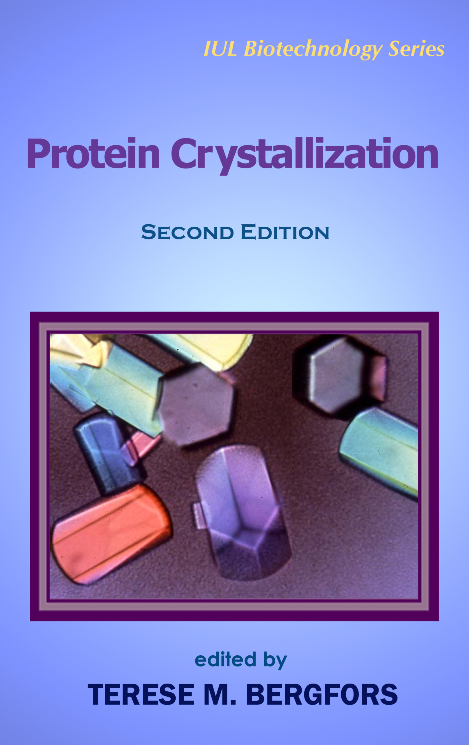
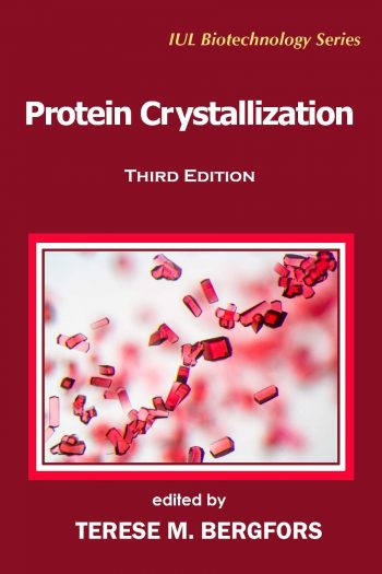
Protein Crystallization
$109.95
IUL Biotechnology Series, 8
by Terese M. Bergfors (Editor)
Edition: Second
Book Details:
- Series: IUL Biotechnology Series
- Volume: 8
- Binding: Hardcover
- Pages: 504
- Dimensions (in inches): 1.20 x 9.75 x 6.50
- Publisher: International University Line
- Publication Date: April 5, 2009
- ISBN: 978-0-9720774-4-6
- Price: $109.95
Second Edition edited by The earliest incarnation of this book was a photocopied manuscript based on students’ questions. I began to compile materials and protocols by way of answering them. The fact that this book is now in its second edition, completely revised and updated, is an evidence of the rapid development and importance of crystallization. <> Acknowledgements
PROTEIN CRYSTALLIZATION
TERESE M. BERGFORS
Contents
Foreword . . . . . vii
Preface . . . . . xix
Contributors . . . . . xxiii
Part I. METHODS . . . . . 1
1. Some Words of Advice from an Old Hand . . . . . 3
Alexander McPherson
2. Rational Selection of Crystallization Techniques . . . . . 11
Joseph R. Luft and George T. DeTitta
2.1. Introduction . . . . . 13
2.2. Some Initial Considerations . . . . . 14
2.2.1. Screening . . . . . 15
2.2.2. Optimization . . . . . 15
2.2.3. The Container . . . . . 15
2.2.4. Temperature . . . . . 6
2.3. Choice of Method . . . . . 17
2.3.1. The Phase Diagram . . . . . 18
2.4. Batch Crystallization Methods . . . . . 19
2.4.1. How to Set Up a Batch Experiment . . . . . 22
2.4.2. Batch Methods That Use Oil . . . . . 23
2.5. Vapor-Diffusion Crystallization Methods . . . . . 23
2.5.1. How to Set Up a Vapor-Diffusion Experiment . . . . . 24
2.5.2. Choice of Reservoir Solution . . . . . 25
2.5.3. PEG Solutions and the Vapor Pressure of Water . . . . . 25
2.5.4. Volatile Additives . . . . . 26
2.5.5. Condensation . . . . . 26
2.5.6. Methods to Prevent Crystal Adhesion . . . . . 27
2.5.7. Containers and Supports for Hanging-Drop Experiments . . . . . 27
2.5.8. Sealants . . . . . 28
2.5.9. Sitting-Drop Vapor-Diffusion Methods . . . . . 29
2.6. Important Variables That Affect Kinetics . . . . . 29
2.6.1. Drop Volume and Shape . . . . . 30
2.6.2. Chemical Concentrations . . . . . 30
2.6.3. Different Chemicals Have Different Rates of Equilibration . . . . . 30
2.6.4. Experiment Drop Volume Ratios . . . . . 31
2.6.5. Drop-to-Reservoir Volume Ratio . . . . . 31
2.6.6 Passive Methods to Regulate the Rate of Vapor-Phase Equilibration . . . . . 32
2.7. Liquid-Diffusion Crystallization Methods . . . . . 32
2.7.1. Dialysis Liquid Diffusion . . . . . 33
2.7.2. How to Set Up a Dialysis Experiment . . . . . 33
2.7.3. Free-Interface Diffusion . . . . . 35
2.7.4. Counterdiffusion . . . . . 35
2.8. Practical Considerations for Crystallization Experiments . . . . . 37
2.8.1. A Chronological Sequence for Hanging-Drop Setups . . . . . 37
2.8.2. Mixing Experiment Drops . . . . . 38
2.9. Some Final Thoughts . . . . . 39
2.10. Summary of Key Points . . . . . 39
Acknowledgments . . . . . 40
Supplies . . . . . 40
References . . . . . 40
3. Automation for Crystallization: Practical Considerations in Choosing a System . . . . . 47
Joseph R. Luft and George T. DeTitta
3.1. Introduction . . . . . 49
3.2. Benefits of Automation . . . . . 50
3.2.1. Reduced Requirements for Protein Sample . . . . . 50
3.2.2. Replication . . . . . 51
3.2.3. Unstable Proteins . . . . . 51
3.2.4. Improved Output . . . . . 51
3.3. Automation for Liquid Handling of the Stock Solutions . . . . . 51
3.3.1. Formulation and Delivery of Cocktail Solutions . . . . . 52
3.3.2. Wash Cycles . . . . . 54
3.4. Automation for Crystallization . . . . . 54
3.4.1. Crystallization Method . . . . . 55
3.4.2. Screening, Optimization, or Both? . . . . . 55
3.4.3. Retrieving Crystals . . . . . 56
3.4.4. Dead, Priming, and Experiment Volumes . . . . . 56
3.4.5. Contact or Noncontact Delivery . . . . . 57
3.4.6. User Interface . . . . . 57
3.4.7. Periodic Maintenance and Quality Control . . . . . 57
3.5. Automation for Imaging the Experimental Outcomes . . . . . 58
3.5.1. Image Quality . . . . . 58
3.6. Automation for Information Management . . . . . 59
3.7. Caveat Emptor (Buyer Beware) . . . . . 60
3.7.1. Excess Capacity . . . . . 60
3.7.2. Personnel Costs . . . . . 60
3.7.3. Market Inquiries . . . . . 61
3.8. Concluding Remarks . . . . . 61
3.9. Summary of Key Points . . . . . 62
Acknowledgments . . . . . 62
References . . . . . 63
4. Oils for Screening and Optimization . . . . . 65
Naomi E. Chayen
4.1. Introduction . . . . . 67
4.2. The Rationale for Crystallization under Oil . . . . . 68
4.2.1. The Microbatch Technique . . . . . 68
4.2.2. Setting up Crystallization Trials by Microbatch . . . . . 68
4.2.3. Crystallization of Membrane Proteins in Oil . . . . . 70
4.2.4. Additional Benefits of Oil . . . . . 70
4.2.5. Harvesting Microbatch Crystals . . . . . 71
4.3. The Effect of Different Oils on Microbatch . . . . . 72
4.3.1. The Use of Different Oils in Screening . . . . . 72
4.3.2. Limitations of Crystallizing under Oil . . . . . 73
4.4. Use of Oils in Optimization . . . . . 73
4.4.1. Control of Nucleation . . . . . 74
4.4.2. Elimination of Surface Contact Leading to Access Nucleation . . . . . 74
4.5. Oil for the Miniaturization and Automation of Crystallization in Gels . . . . . 76
4.6. The Use of Oil for Controlling the Rate of Vapor-Diffusion Trials . . . . . 76
4.7. Conversion between Vapor Diffusion and Batch/Microbatch . . . . . 77
4.8. Summary . . . . . 79
References . . . . . 80
5. Crystallization in Gels and Capillaries . . . . . 83
Juan M. García-Ruiz
5.1. Why Use Gels and Capillaries for Protein Crystallization? . . . . . 85
5.2. Is Mass Transport by Diffusion Slower than Mass Transport by Convection? . . . . . 86
5.3. Is a Diffusive Environment Better than a Convective One for Obtaining High-Quality Crystals? . . . . . 87
5.4. Is There Convection in Vapor-Diffusion Experiments? . . . . . 88
5.5. How Can Convection Be Reduced in Crystallization Experiments? . . . . . 88
5.6. Which Gels Should Be Used? . . . . . 89
5.7. Is It Better to Use Gels or Capillaries for Reducing Convection? . . . . . 89
5.8. Can Gels and Capillaries Be Used with Classical Crystallization Techniques? . . . . . 90
5.9. How Does the Counterdiffusion Technique Work? . . . . . 90
5.10. Isn’t a Test Tube Too Large for the Small Amount of Proteins Usually Available? . . . . . 91
5.11. Is It Difficult to Fill Such a Small Capillary? . . . . . 92
5.12. What Kind of Capillaries Should Be Used for Counterdiffusion? . . . . . 92
5.13. Is It Possible to Cryoprotect the Crystals in the Capillaries? . . . . . 93
References . . . . . 93
6. Seeding . . . . . 95
Aengus Mac Sweeney and Allan D’Arcy
6.1. Introduction . . . . . 97
6.2. Streak Seeding . . . . . 99
6.3. Solution Microseeding . . . . . 101
6.3.1. Preparing a Microseed Stock . . . . . 101
6.3.2. Determining the Best Conditions for Solution Microseeding . . . . . 103
6.4. Macroseeding . . . . . 104
6.4.1. Obtaining the Best Possible Seed Crystals . . . . . 105
6.4.2. Determining the Crystal Solubility . . . . . 105
6.4.3. Determining Appropriate Crystal-Growth Conditions . . . . . 106
6.4.4. Washing and Transferring Crystals . . . . . 106
6.4.5. Feeding . . . . . 107
6.5. Seeding When Using Additives . . . . . 107
6.6. Automation and Microseeding . . . . . 108
6.7. Heterogeneous Seeding . . . . . 108
6.8. Summary . . . . . 111
6.9. Summary of Key Points . . . . . 112
References . . . . . 113
7. Heavy-Atom Derivatization . . . . . 115
Zbigniew Dauter and Mirosława Dauter
7.1. Why Heavy Atoms? . . . . . 117
7.2. Precautions . . . . . 119
7.3. Types of Heavy Atoms . . . . . 120
7.3.1. Classic Heavy-Metal Compounds . . . . . 120
7.3.2. Mercurials . . . . . 122
7.3.3. Lanthanides . . . . . 123
7.3.4 Metal Clusters . . . . . 123
7.3.5. Noble Gases . . . . . 124
7.3.6. Halides . . . . . 124
7.3.7. Iodination of Aromatics . . . . . 125
7.3.8. Rapid Soaks . . . . . 126
7.4. Confirming Derivatization . . . . . 127
7.4.1. Quick, Low-Resolution Data Collection . . . . . 127
7.4.2. Mass Spectrometry . . . . . 127
7.4.3. microPIXE . . . . . 128
7.4.4. X-Ray Absorption Spectroscopy . . . . . 128
7.4.5. Native Gel Shift Assay . . . . . 129
7.5. Radiation Damage . . . . . 129
7.6. Final Remarks . . . . . 130
References . . . . . 130
8. Preparation and Crystallization of Selenomethionine Protein . . . . . 135
Anna M. Larsson
8.1. Why Use Selenomethionine (SeMet)? . . . . . 137
8.2. When to Choose SeMet . . . . . 138
8.2.1. Methionine Content . . . . . 138
8.2.2. Expression Hosts . . . . . 139
8.2.3. Incorporation Analysis . . . . . 139
8.3. Other Related Approaches . . . . . 141
8.3.1. Selenocysteine . . . . . 141
8.3.2. Sulfur-SAD Phasing . . . . . 141
8.4. Properties of SeMet . . . . . 142
8.4.1. How to Avoid Oxidation . . . . . 142
8.4.2. How to Induce Oxidation and Why . . . . . 142
8.4.3. Sensitivity to Radiation Damage . . . . . 143
8.4.4. Data Collection Strategies . . . . . 143
8.4.5. L– or D,L-Selenomethionine . . . . . 143
8.4.6. Warning . . . . . 143
8.5. How to Express SeMet Protein . . . . . 144
8.5.1. Expression of SeMet-Labeled Protein in Escherichia coli . . . . . 144
8.5.2. Autoinduced Expression of SeMet-Labeled Protein in Escherichia coli . . . . . 145
8.5.3. Expression of SeMet-Labeled Protein in Pichia pastoris . . . . . 145
8.6. How to Purify and Crystallize SeMet Protein . . . . . 146
8.6.1. Purification . . . . . 146
8.6.2. Crystallization . . . . . 146
8.7. Summary . . . . . 147
References . . . . . 148
Appendix 8.1. Reagents for the Media . . . . . 151
9. Crystal Handling for Cryogenic Data Collection . . . . . 155
Elspeth F. Garman
9.1. Introduction . . . . . 157
9.2. Protocol for Finding Crystallization Conditions for Cryocrystallography . . . . . 162
9.3. Practical Tips for Crystal Transfer and Handling . . . . . 168
9.4. Methods for Introducing the Crystal into the Cryosolution . . . . . 169
9.4.1. Soaking . . . . . 169
9.4.2. Exchanging Mother Liquors . . . . . 169
9.4.3. Osmolarity Matching . . . . . 170
9.5. Postcrystallization Treatments . . . . . 170
9.5.1. Dry Mounting . . . . . 170
9.5.2. Annealing . . . . . 170
9.6. Summary of Key Points . . . . . 171
References . . . . . 172
PART II. TOOLS and STRATEGIES . . . . . 175
10. Interpretation of the Crystallization Drop Results . . . . . 177
Johan Philip Zeelen
10.1. Introduction . . . . . 179
10.2. The Stereomicroscope . . . . . 180
10.2.1. Bright- and Dark-Field Illumination . . . . . 180
10.2.2. Polarizers and Birefringence . . . . . 181
10.3. Optical Methods for Distinguishing Crystals of Protein, Salt, and Detergent . . . . . 182
10.4. Examination of the Crystallization Experiments . . . . . 184
10.5. Scoring . . . . . 185
10.6. Interpretation of the Drop Phenomena . . . . . 187
References and Further Reading . . . . . 193
11. Mass Spectrometry Applications in Protein Crystallography . . . . . 195
Sharon X. Gao and Marie Zhang
11.1. Introduction . . . . . 197
11.2. Mass Spectrometry Instrumentation . . . . . 198
11.2.1. Molecular Mass . . . . . 198
11.2.2. MALDI-MS . . . . . 199
11.2.3. ESI-MS . . . . . 202
11.2.4. Matrix-Free Desorption or Ionization on Porous Silicon (DIOS) . . . . . 204
11.3. Applications of MS in Protein Structure Determination . . . . . 204
11.3.1. High-Throughput MS Methods for Monitoring Protein Purity . . . . . 205
11.3.2. Verification of Protein Primary Structure . . . . . 205
11.3.3. Characterization of Posttranslational Modifications . . . . . 207
11.3.4. Detection of Heavy Atoms for Phasing . . . . . 209
11.3.5. Limited Proteolysis MS for Domain Mapping . . . . . 210
11.3.6. Large-Scale Limited Proteolysis and Crystallization Studies . . . . . 211
11.3.7. Probing Higher-Order Protein Structure with MS . . . . . 211
11.3.8. Analysis of the Content of Protein Crystals by MS . . . . . 212
11.4. Concluding Remarks . . . . . 213
References . . . . . 214
12. Dynamic Light Scattering . . . . . 221
Ulf Nobbmann and Terese Bergfors
12.1. Introduction . . . . . 223
12.1.1. How Does DLS Work? . . . . . 226
12.1.2. Relationship between Hydrodynamic Radius and Mass . . . . . 229
12.1.3. How Is the Dispersity Profile Obtained? . . . . . 230
12.2. Practical How-To . . . . . 233
12.2.1. Comparison with the Native PAGE . . . . . 233
12.2.2. Sample Preparation: Volume and Concentration . . . . . 234
12.2.3. Filtration . . . . . 235
12.2.4. Temperature . . . . . 235
12.2.5. More Things to Try: Increase the Ionic Strength . . . . . 236
12.2.6. Change the pH . . . . . 237
12.2.7. Increase Protein Concentration . . . . . 37
12.2.8. Additives . . . . . 238
12.3. Thermal Stability Measurements with DLS . . . . . 240
12.4. Data Interpretation . . . . . 241
12.5. Some Recommendations for Handling Your Protein Based on Its DLS Profile . . . . . 242
12.5.1. Monodisperse Samples (approximately 20% or less) . . . . . 242
12.5.2. Monodisperse Samples (approximately 30% or less) . . . . . 242
12.5.3. Bimodal and Polydisperse Samples . . . . . 243
12.6. Summary of Key Points . . . . . 243
References . . . . . 244
13. Improving Protein Crystallizability by Modifications and Engineering . . . . . 247
Xiayang Qiu and Cheryl A. Janson
13.1. Introduction . . . . . 249
13.2. Ligand Additions and Complexes . . . . . 250
13.2.1. Cofactors, Substrates, Inhibitors, and Other Ligands . . . . . 250
13.2.2. Protein–Protein and Protein–Nucleic Acid Complexes . . . . . 251
13.2.3. Complexes with Antibody Fragments . . . . . 251
13.3. Chemical and Biochemical Modifications . . . . . 252
13.3.1. Chemical Modification . . . . . 252
13.3.2. Biochemical Modification . . . . . 253
13.4. DNA-Construct Modification . . . . . 254
13.4.1. Affinity Tags and Protease Removal Sites . . . . . 254
13.4.2. Defining Domains for Construct Design . . . . . 255
13.4.3. Surface Mutations to Improve Solution Properties . . . . . 257
13.4.4. Surface Mutations to Facilitate Crystal Packing . . . . . 258
13.4.5. Metal–Ion-Mediated Crystallization . . . . . 260
13.5. Concluding Remarks . . . . . 262
References . . . . . 262
14. Handling the Protein Sample . . . . . 267
Terese Bergfors
14.1. Characterization of the Protein . . . . . 270
14.2. Concentrating the Protein . . . . . 271
14.3. Storage of the Protein . . . . . 273
14.3.1. Freezing . . . . . 273
14.3.2. Lyophilization . . . . . 276
14.3.3. Ammonium Sulfate Precipitation . . . . . 277
14.3.4. Heat Shock . . . . . 277
14.4. Buffer Choice: Implications for Screening . . . . . 279
14.5. Other Components in the Start Buffer . . . . . 282
14.5.1. Water . . . . . 282
14.5.2. Sodium Azide . . . . . 283
14.5.3. Reducing Agents . . . . . 284
14.6. Buffer Choice: Implications for Screening . . . . . 287
14.7. Other Components in the Start Buffer . . . . . 289
Acknowledgments . . . . . 289
References . . . . . 290
15. Two Approaches for Initial Screening: Evolution and Intelligent Design . . . . . 293
Janet Newman
15.1. Introduction . . . . . 295
15.2. Why Design a New Screen? . . . . . 296
15.2.1. Learning from the Past: Evolution (JCSG+) . . . . . 297
15.2.2. Intelligent Design (PACT) . . . . . 298
15.2.3. Does Limited Screening Work? . . . . . 302
15.3. Screening to Ease Optimization . . . . . 303
15.4. Designing Your Own Screen . . . . . 306
15.5. Summary of Key Points . . . . . 307
Supplies . . . . . 307
References . . . . . 307
16. Strategies for Protein Crystallization Screening . . . . . 309
Bernhard Rupp
16.1. Introduction . . . . . 311
16.1.1. Protein Crystals . . . . . 312
16.1.2. Basics of Protein Crystallization . . . . . 313
16.1.3. Beating the Odds . . . . . 313
16.2. Designing Your Protein . . . . . 314
16.2.1. Constructs and Tags . . . . . 314
16.2.2. Batch Variation and Contaminants . . . . . 315
16.2.3. Protein Concentration . . . . . 315
16.2.4. Purity, Freshness, and Conformational State . . . . . 316
16.2.5. Buffers, Salts, and Additives . . . . . 316
16.2.6. Temperature . . . . . 316
16.3. Choice of Crystallization Technique . . . . . 317
16.4. Crystallization Screening . . . . . 318
16.4.1. Crystallization As a Multivariate Sampling Problem . . . . . 319
16.4.2. Factorial Designs . . . . . 320
16.4.3. Sparse-Matrix Sampling . . . . . 321
16.4.4. Random Sampling and Number of Experiments . . . . . 322
16.4.5. Screening toward Optimization . . . . . 322
16.4.6. Practical Two-Tiered Approaches . . . . . 323
16.5. Robotics in the Academic Laboratory . . . . . 324
16.5. Summary of Key Points . . . . . 326
References . . . . . 327
17. Additives and Microcalorimetric Approaches for Optimization of Crystallization . . . . . 331
Joanne I. Yeh
17.1. Introduction . . . . . 333
17.2. Classes of Additives . . . . . 334
17.2.1. Cosmotropes . . . . . 334
17.2.2. Cations . . . . . 335
17.2.3. Polyols and Sugars . . . . . 335
17.2.4. Amino Acids . . . . . 336
17.2.5. Linkers . . . . . 336
17.2.6. Cofactors, Ligands, Substrates (Effectors) . . . . . 337
17.2.7. Reducing Agents and Chelators . . . . . 337
17.2.8. Chaotropes . . . . . 339
17.2.9. Surfactants . . . . . 339
17.3. What Concentration of Additive to Use . . . . . 341
17.4. Calorimetry Studies on Additives . . . . . 342
17.4.1. Instrumentation . . . . . 344
17.4.2. ITC and DSC Results . . . . . 346
17.5. Summary . . . . . 347
References . . . . . 347
PART III. LABORATORY EXERCISES . . . . . 351
LE. Laboratory Exercises . . . . . 353
LE1. Setting Up Microbatch Trials . . . . . 355
Naomi E. Chayen
LE2. Limiting the Amount of Nucleation and Enhancing the Reproducibility of Experiments by Filtration . . . . . 359
Naomi E. Chayen
LE3. Containerless Crystallization . . . . . 363
Naomi E. Chayen
LE4. Insertion of Oil Barriers to Slow Down Vapor-Diffusion Experiments . . . . . 367
Naomi E. Chayen
LE5. Improving Crystal Quality by Separating Nucleation and Growth in Hanging Drops . . . . . 371
Naomi E. Chayen
LE6. Seeding with Lysozyme . . . . . 375
Aengus Mac Sweeney
LE7. Studying the Influence of Supersaturation and Supersaturation Rate with the Crystallization Mushroom . . . . . 387
Juan Manuel García-Ruiz and Luis A. González-Ramírez
LE8. Capillary Counterdiffusion Experiments with Granada Crystallization Boxes . . . . . 395
Juan Manuel García-Ruiz and Luis A. González-Ramírez
LE9. Evaluation of Cryoprotectant Solutions . . . . . 401
Elspeth F. Garman
LE10. Optimization Experiment with Additives . . . . . 405
Terese Bergfors
LE11. Comparing Salt and Protein Crystals by Two Methods . . . . . 409
Terese Bergfors
PART IV. A–Z . . . . . 415
A–Z . . . . . 417
Index . . . . . 427
Foreword
This second edition of Protein Crystallization includes all the important methods, tools, and strategies that are presently used for crystallizing proteins. The result is a most impressive book—almost completely new as compared to the first edition. As with the first edition, the emphasis is on the practical procedures for performing the work in the laboratory. It will be an invaluable guide at the laboratory bench both for beginners and more experienced crystallizers of biological macromolecules.
I have known Terese Bergfors since she came to our department in 1984. She has become one of the leading experts in the field of protein crystallization and her contributions are invaluable for her colleagues at our institute as well as for many crystallographers in other departments. She is an enthusiastic teacher and frequently teaches courses in protein crystallization here in Sweden and abroad. Terese Bergfors has done an excellent job of persuading well-known fellow scientists in her field to share their knowledge and experience on specific
crystallization topics as authors for the different chapters of this book.
In my view, this book constitutes perfect evidence that protein crystallization should now be considered a science rather than an art. However, as is strongly emphasized in the chapter by Joe Luft and George DeTitta, this presupposes that all the factors that influence crystallization are well controlled, even on a detailed level.
The fact that this book is now in its second edition shows how rapidly our knowledge about protein crystallization is growing. The field has changed tremendously since its early days. In the beginning of the l960s, soon after the structures of myoglobin and hemoglobin were solved in Cambridge, our group in Uppsala started the structural determination of the enzyme human carbonic anhydrase. We purified the protein in enormous volumes from liters and liters of blood. The blood came from the University hospital after it had become too
old to be used for blood transfusions. The crystallizations were done in dialysis bags containing milliliters of protein solution, a huge difference to the nanoliter volumes used today.
In his chapter, Alexander McPherson concludes his thoughtful words of advice with the remark that “further advances in the field of protein crystallization are essential to greater progress in structural and molecular biology.” He encourages us all to contribute with new ideas, novel approaches, etc. Ideally this book will be a springboard for a development in this direction, as well as a great stimulus and guide for all protein crystallographers.
Professor Emeritus Bror Strandberg, Uppsala University
Member of Sir John Kendrew’s research team when determining the first
three-dimensional structure of a protein (myoglobin) almost fifty years ago.
Preface to the Second Edition
The accumulated knowledge on practical aspects of protein crystallization is scattered in many different sources or in the form of local lab lore. The value of a book on crystallization is that it gathers the material and organizes it in one place. The authors of the chapters are leading experts in their techniques and areas and many of them are also active as teachers. My goal as editor has been to provide an overall unity and cohesion to the 17 chapters. The chapter styles vary—some are written as reviews and others are informal and practical. There
are frequent cross-references between chapters to help the reader find additional information on a topic. Some overlap of material has been permitted intentionally for the benefit of the readers who will be dipping into the book, rather than reading it from cover to cover.
If you already have the first edition of Protein Crystallization, why should you buy this second edition? You will find that almost all of the material is new. The first edition was published in 1999, when high-throughput consortia were in their infancy. In the few years since then, high-throughput methods have contributed to the miniaturization and automation of crystallization experiments as well as a vast pool of data on crystallization screening. New strategies, based on these data, are taken up in two separate chapters by Newman and Rupp. Automation is becoming affordable even for small laboratories and its importance will grow. Other chapters new for this edition cover guidelines for choosing automation for crystallization. These have been written by Luft, DeTitta, and
Rupp, based on their expertise from high-throughput laboratories.
Also new for this edition are chapters on mass spectrometry (Gao and Zhang), microcalorimetry (Yeh), counterdiffusion (García-Ruiz), heavy-atom derivatization (Dauter and Dauter), selenomethionine proteins (Larsson) and engineering proteins to make them more amenable to crystallization (Qiu and Janson). Zeelen has contributed again from his library of crystallization pictures so that this section now contains four pages of color plates. Interpretation of the phenomena in the crystallization drop is an area in which beginners (and automated visual recognition systems) have great difficulty. To my mind it is the ability to recognize promising leads and knowing what to do with them that constitutes the proverbial green thumb in crystallization. Study the photographs and compare your drops with them—the time will be well invested.
McPherson opens the book once again with some basic advice. Seeding is reviewed by Mac Sweeney and D’Arcy and an extensive seeding exercise is included. The chapter on crystallization methods is in much greater depth and includes excellent illustrations and phase-diagram explanations by Luft. For the chapter on dynamic light scattering, Nobbmann explains the theoretical principles and I cover the practical issues. Chayen, Garman, and I have updated and expanded the chapters we wrote for the first edition. Membrane proteins are not
treated as a separate chapter in this edition although they are covered within chapters as appropriate. This is because there are now several books specifically on the crystallization of membrane proteins, for example, So Iwata’s Methods and Results in Crystallization of Membrane Proteins, also published by International University Line.
The laboratory exercises are grouped together for this edition in a separate section to facilitate their use in courses. Many new ones have been added. The exercises have been tested in the “Practical Protein Crystallization” course that I have been running since 1994 at Uppsala University, as well as courses elsewhere. Your feedback on these, or any aspect of the book, is greatly welcomed—please send any questions or suggestions to me, [email protected].
The authors generously took their time to contribute to this book. I am indebted to them for their efforts. It has been a privilege to work with them and I have learned many things from the numerous discussions we shared this past year.
I thank my colleagues who read the chapters and exercises and gave me valuable input. Some of my test readers deserve special mention: Anna Jansson, Bror Strandberg, Lars Liljas, Wojciech Krajewski, Gerard Kleywegt (all from Uppsala University), Jonas Vasur (Swedish Agricultural University), Joe Ng (University of Alabama in Huntsville), and Inés Muñoz (CNIO, Madrid). Matti Nikkola (Karolinska Institutet, Stockholm) and Christina Skans extricated me from some murky linguistic cul-de-sacs with their impressive knowledge of
English. Mark Harris (Uppsala University) gave me invaluable help with the figures and his patience in explaining PhotoShop to me over and over verges on the saintly. Professor Alwyn Jones (Uppsala University) generously allowed me to devote myself to the book without interruption. Dr. Igor Tsigelny (International University Line) is editor of the series in which this book is a part. I am grateful to him for the opportunity to turn my photocopied papers into a book that has now reached its second edition. I thank Stefan Odestedt for sorting out the mysteries of Microsoft Excel for me and his moral support in general. Finally, I extend my deepest appreciation to my daughter, Carmen, for her forbearance yet once again during my preoccupation with this book.
Uppsala, Sweden
March 2009
Terese Bergfors




Dr. Herbert A. Hauptman –
Nobel Laureate in Chemistry
Why is crystallization important? At different times, different aspects of protein crystallography have assumed the role of bottleneck. Thus at one time the bottleneck consisted of the lack of a methodology for structure determination, even if a sufficient number of diffraction intensitities were available for structure solution. With the advent of powerful methods for structure solution, the bottleneck shifted to the difficulty in obtaining crystals in sufficient number and of suitable quality to do the diffraction experiment. The present book is dedicated to the problem of describing current methods for protein crystallization and describing them as clear laboratory protocols.
Dr. William L. Duax –
American Crystallographic Association, Chief Executive Officer
This is an essential handbook for anyone engaged in crystallization of macromolecules. It is exceptionally well organized and illustrated and has contributions from all the leaders in what continues to be a challenging and critically important field.