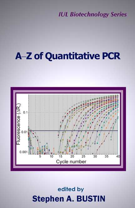
A-Z of Quantitative PCR
$119.95
IUL Biotechnology Series, 5
by Stephen A. Bustin (Editor)
Edition: First
Print: Third
Book Details:
- Series: IUL Biotechnology Series
- Volume: 5
- Binding: Hardcover
- Pages: 912
- Dimensions (in inches): 1.75 x 9.50 x 6.50
- Publisher: International University Line
- Publication Date: August, 2004
- ISBN: 0-9636817-8-8
- Price: $109.95
This is not just a cook book for real-time quantitative PCR (qPCR). Admittedly, there are lots of recipes from distinguished contributors and I have attempted to collect, sift through and rationalize the vast amount of information that is available on this subject. And yes, this book was conceived as a comprehensive hands-on manual to allow both the novice researcher and the expert to set up and carry out qPCR assays from scratch. However, this book also sets out to explain as many features of qPCR as possible, provide alternative viewpoints and methods and, perhaps most importantly, aims to stimulate the researcher into generating, interpreting and publishing data that are reproducible, reliable, and biologically meaningful. Stephen A. Bustin
A-Z of Quantitative PCR
edited by
Stephen A. Bustin
Contents
Preface xxi
List of Contributors xxiii
Acronims and Abbreviations xxvii
Part I. OVERVIEWS 1
1. Quantification of Nucleic Acids by PCR 3
Stephen A. Bustin
1.1. Introduction 5
1.1.1. PCR Characteristics 6
1.2. Conventional Quantitative PCR 8
1.2.1. Concepts 10
1.2.2. Limitations 12
1.2.3. Alternatives 13
1.3. Real-Time Quantitative PCR 15
1.3.1. Uses 16
1.3.2. Microdissection 19
1.3.3. Limitations 22
1.3.4. PCR 22
1.3.5. RT-PCR 23
1.4. Outlook 26
1.5. Conclusion 29
2. Real-Time RT-PCR: What Lies Beneath the Surface 47
Jonathan M. Phillips
2.1. Introduction 49
2.2. What is RT-PCR? 50
2.2.1. Reverse Transcription and RT Enzymes 52
2.2.2. What is Quantitative RT-PCR? 57
2.2.3. Real-Time RT-PCR 58
2.2.4. Reaction Controls (IPCs) 58
2.2.5. Reporter Technologies 60
2.3. Things That Influence RT-PCR 61
2.3.1. Why Commercial Kits? 62
2.3.2. Divalent Metal Concentration 64
2.3.3. Primer Concentration 65
2.3.4. Probe Concentration 66
2.3.5. Reverse Transcription Conditions 67
2.4. Synthetic Molecules 70
2.4.1. Substituted Primers and Probes 70
2.4.2. Synthetic RNA Controls 71
2.5. A Word about DNA Polymerases 73
2.5.1. DNA Dependent DNA Polymerases 73
2.5.2. RNA Dependent DNA Polymerases 74
2.6. Tips and Tricks 75
2.6.1. Probes 75
2.6.2. The Right Enzyme for the Job 77
2.7. Buffers 78
2.8. Concluding Remarks 78
3. Quantification Strategies in Real-Time PCR 87
Michael W. Pfaffl
3.1. Introduction 89
3.2. Markers of a Successful Real-Time RT-PCR Assay 90
3.2.1. RNA Extraction 90
3.2.2. Reverse Transcription 91
3.2.3. Comparison of Real-Time RT-PCR with Classical Endpoint Detection Method 93
3.2.4. Chemistry Developments for Real-Time RT-PCR 94
3.2.5. Real-Time RT-PCR Platforms 94
3.2.6. Quantification Strategies in Kinetic RT-PCR 95
3.2.7. Advantages and Disadvantages of External Standards 100
3.2.8. Real-Time PCR Amplification Efficiency 102
3.2.9. Data Evaluation 105
3.3. Automation of the Quantification Procedure 106
3.4. Normalization 108
3.5. Statistical Comparison 111
3.6. Conclusion 112
PART II. BASICS 121
4. Good Laboratory Practice! 123
Stephen A. Bustin and Tania Nolan
4.1. Introduction 125
4.2. General Precautions 126
4.2.1. Phenol 127
Emergency procedures in case of skin contact 128
4.2.2. Liquid Nitrogen (N2) 129
4.2.3. Waste Disposal 130
4.3. Equipment 131
4.3.1. Electrophoresis 131
4.3.2. Freezer 131
4.3.3. UV Transilluminators 131
4.3.4. Micropipettes 132
4.3.5. Gloves 134
4.3.6. Eye Protection 135
4.3.7. Legal Information 136
5. Template Handling, Preparation, and Quantification 141
Stephen A. Bustin and Tania Nolan
5.1. Introduction 143
5.1.1. General Precautions 144
5.2. DNA 146
5.2.1. Preanalytical Steps 146
5.2.2. Sample Collection 150
5.2.3. Disruption 151
5.2.4. Purification 154
5.2.5. Long-Term Storage 159
5.3. RNA 159
5.3.1. Preanalytical Steps 160
5.3.2. General Considerations 161
5.3.3. Tissue Handling and Storage 163
5.3.4. Disruption/Homogenization 165
5.3.5. RNA Extraction 173
5.3.6. Simultaneous DNA Extraction 180
5.3.7. DNA Contamination 182
5.3.8. Preparation of RNA from Flow Cytometrically Sorted Cells 183
5.3.9. Extraction from Formalin-Fixed and Paraffin-Embedded Biopsies 184
5.3.10. Specialized Expression Analysis 187
5.4. Quantification of Nucleic Acids 188
5.4.1. Absorbance Spectrometry 188
5.4.2. Fluorescence 190
5.4.3. Purity 190
5.4.4. Quantification of RNA 191
6. Chemistries 215
Stephen A. Bustin and Tania Nolan
6.1. Introduction 217
6.2. Fluorescence 221
6.2.1. Fluorophores 222
6.2.2. Quenchers 226
6.3. Nonspecific Chemistries 228
6.3.1. DNA Intercalators 228
6.3.2. Advantages 229
6.3.3. Disadvantages 231
6.3.4. Quencher-Labeled Primer (I) 234
6.3.5. Quencher-Labeled Primer (II) 234
6.3.6. LUX™ Primers 235
6.3.7. Amplifluor™ 236
6.4. Specific Chemistries 239
6.4.1. Advantages 240
6.4.1. Disadvantages 240
6.5. Linear Probes 241
6.5.1. ResonSense® and Angler® Probes 241
6.5.2. HyBeacons™ 242
6.5.3. Light-up Probes 243
6.5.4. Hydrolysis (TaqMan®) Probes 244
6.5.5. Lanthanide Probes 246
6.5.6. Hybridization Probes 249
6.5.7. Eclipse™ 249
6.5.8. Displacement Hybridization/Complex Probe 250
6.6. Structured Probes 251
6.6.1. Molecular Beacons 253
6.6.2. Scorpions™ 259
6.63. Cyclicons™ 261
6.7. Future Technology 263
6.7.1. Nanoparticle Probes 263
6.7.2. Conjugated Polymers And Peptide Nucleic Acid Probes 263
7. Primers and Probes 279
Stephen A. Bustin and Tania Nolan
7.1. Introduction 281
7.1.1. Hybridization 283
7.2. Probe Design 288
7.3. Hydrolysis Probes 290
7.3.1. Gene Expression Analysis 290
7.3.2. SNP/Mutation Analysis 292
7.4. Hybridization Probes 293
7.4.1. Gene Expression Analysis 293
7.4.2. SNP/Mutation Analysis 294
7.5. Molecular Beacons 294
7.5.1. Gene Expression Analysis 295
7.5.2. SNP/Mutation Analysis 296
7.6. Scorpions™ 296
7.6.1. Gene Expression Analysis 297
7.6.2. SNP/Mutation Analysis 299
7.7. Probe Storage 299
7.8. Primer Design 299
7.9. Amplifluor™ Primers 303
7.10. LUX™ Primers 304
7.11. Oligonucleotide Purification 305
7.12. Recommended Storage Conditions 307
7.13. Example of Primer Design 308
7.14. Nucleic Acid Analogues 311
7.14.1. Peptide Nucleic Acids (PNA) 313
7.14.2. PNA Probe Characteristics 315
7.14.3. Locked Nucleic Acids LNA™ 317
7.14.4. Modified Bases: Super A™, G™, and T™ 318
7.14.5. Minor Groove Binding Probes 319
8. Instrumentation 329
Stephen A. Bustin and Tania Nolan
8.1. Introduction 331
8.1.1. The Principle 332
8.1.2. Excitation Source 333
8.1.3. Filters 335
8.1.4. Photodetectors 337
8.1.5. Sensitivity 339
8.1.6. Dynamic Range 340
8.1.7. Linearity 340
8.2. Real-Time Instruments 341
8.2.1. ABI Prism® 345
8.2.2. Bio-Rad Instruments 346
8.2.3. Stratagene’s Instruments 348
8.2.4. Corbett Research Rotor-Gene RG-3000 350
8.2.5. Roche Applied Science 353
8.2.6. Techne Quantica 355
8.2.7. Cepheid Smart Cycler® 356
8.3. Outlook 355
9. Basic RT-PCR Considerations 359
Stephen A. Bustin and Tania Nolan
9.1. Introduction 361
9.2. Total RNA vs. mRNA 364
9.3. cDNA Priming 364
9.3.1. Random Primers 365
9.3.2. Oligo-dT 366
9.3.3. Target-Specific Primers 366
9.4. Choice of Enzyme 366
9.4.1. RT Properties 367
9.4.2. AMV-RT 370
9.4.3. MMLV-RT 371
9.4.4. DNA-Dependent DNA Polymerases 372
9.4.5. Omniscript/Sensiscript 372
9.5. RT-PCR 372
9.5.1. Two-Enzyme Procedures: Separate RT and PCR Enzymes 373
9.5.2. Single RT and PCR Enzyme 374
9.5.3. Problems with RT 375
9.6. One-Enzyme/One-Tube RT-PCR Protocol 376
9.6.1. Preparations 376
9.6.2. Primers and Probes 376
9.6.3. RT-PCR Enzyme 377
9.6.4. RT-PCR Solutions 377
9.6.5. Preparation of Master Mix 377
9.6.6. Preparation of Standard Curve 378
9.6.7. Template Reaction 380
9.6.8. Troubleshooting 381
9.7. Two-Enzyme/Two-Tube RT-PCR Protocol 382
9.7.1. RT-PCR Enzymes 382
9.7.2. RT-PCR Solutions 382
9.7.3. Preparation of Master Mix 382
9.7.4. Preparation of Standard Curve 383
9.7.5. Unknown Template Reaction 385
9.7.6. Troubleshooting 386
10. The PCR Step 397
Stephen A. Bustin and Tania Nolan
10.1. Introduction 399
10.2. Choice of Enzyme 400
10.3. Thermostable DNA Polymerases 401
10.3.1. Fidelity 406
10.3.2. Processivity and Elongation Rates 406
10.3.3. Thermostability 407
10.3.4. Robustness 407
10.4. To UNG or not to UNG 410
10.5. Hot Start PCR 411
10.6. PCR Assay Components 413
10.6.1. Enzyme Concentration 413
10.6.2. Mg2+ Concentration 414
10.6.3. Primers 414
10.6.4. dNTPs 415
10.6.5. Template 416
10.6.6. Inhibition of PCR by RT Components 417
10.6.7. Water 417
10.7. Reaction Conditions 417
10.7.1. Denaturation Temperature 418
10.7.2. Annealing Temperature 418
10.7.3. Polymerization Temperature 418
10.7.4. Reaction Times 419
10.7.5. Multiplexing 419
10.7.6. Additives 419
10.8. PCR Protocols for Popular Assays 422
10.8.1. Preparations 423
10.8.2. Double Stranded DNA Binding Dye Assays 424
10.8.3. Hydrolysis (TaqMan) Probe Reaction 426
10.8.4. Molecular Beacon Melting Curve to Test Beacon and Scorpion Assays 429
10.8.5. Molecular Beacon/Scorpion Reaction 430
10.9. General Troubleshooting 431
11. Data Analysis and Interpretation 439
Stephen A. Bustin and Tania Nolan
11.1. Introduction 441
11.2. Precision, Accuracy, and Relevance 442
11.3. Quantitative Principles 444
11.4. Effect of Initial Copy Numbers 446
11.5. Monte Carlo Effect 447
11.6. Amplification Efficiency 448
11.7. Relative, Comparative or Absolute Quantification 449
11.8. Absolute Quantification 450
11.9. Standard Curves 451
11.9.1. Recombinant DNA 454
11.9.2. Genomic DNA 455
11.9.3. SP6 or T7-Transcribed RNA 456
11.9.4. Universal RNA 456
11.9.5. Sense-Strand Oligonucleotides 457
11.10. Relative Quantification 458
11.11. Normalization 460
11.11.1. Tissue Culture 461
11.11.2. Nucleated Blood Cells (NBC) 462
11.11.3. Solid Tissue Biopsies 462
11.11.4. Cell Number 463
11.11.5. Total RNA 463
11.11.6. DNA 464
11.11.7. rRNA 464
11.12. Reference Genes (Housekeeping Genes) 465
11.13. Basic Statistics 467
11.13.1. Data Presentation 469
11.13.2. Mean and Median 469
11.13.3. Standard Deviation 470
11.13.4. Plots 470
11.13.5. Relative (Receiver) Operating Characteristics 471
11.13.6. Probability 473
11.13.7. Parametric and Nonparametric Tests 475
11.14. Conclusion 481
12. The qPCR Does Not Work? 493
Stephen A. Bustin and Tania Nolan
12.1. Introduction 495
12.2. Problem: What Is a Perfect Amplification Plot? 496
12.3. Problem: Too Much Target 498
12.9.1. Solution 499
12.4. Problem: Amplification Plot Is not Exponential 499
12.4.1. Solution 500
12.5. Problem: Duplicates Give Widely Differing Cts 500
12.5.1. Solution 502
12.6. Problem: No Amplification Plots 502
12.6.1. Solution 502
12.7. Problem: The Probe Does not Work! 506
12.7.1. Solution 510
12.8. Problem: The Data Plots Are Very Jagged 511
12.8.1. Solution 511
12.9. Problem: The Amplification Plot for the Standard Curve Looks Great BUT…………… 512
12.9.1. ……..The Gradient of the Standard Curve Is Greater Than -3.3 514
12.9.2. ……..The Standards Aren’t Diluting! 515
12.9.3. ……..Using SYBR Green the Gradient of the Standard Curve Is Less Than -3.3 517
12.9.4. ……..Using a Sequence Specific Oligonucleotide Detection System the Gradient of the
Standard Curve Is Less Than -3.3 518
12.10. Problem: The Amplification Plots Are Strange Wave Shapes 521
12.10.1. Solution 522
12.11. Problem: The Amplification Plot Goes Up, Down and All Around 523
12.11.1. Solution 523
PART III. SPECIFIC APPLICATIONS 525
13. Getting Started—The Basics of Setting up a qPCR Assay 527
Tania Nolan
13.1. Introduction 529
13.2. Optimization 531
13.3. Primer and Probe Optimization Protocol 532
13.4. Optimization of Primers Concentration Using SYBR Green I 534
13.5. SYBR Green 1 Optimization Data Analysis 535
13.6. Examination of the Melting Curve 535
13.7. Optimization of Primer Concentration Using Fluorescent Probes 537
13.8. Molecular Beacon Melting Curve 537
13.9. Primer Optimization Reactions in Duplicate 538
13.10. Primer Optimization Data Analysis 539
13.11. Optimization of Probe Concentration 539
13.12. Probe Optimization Data Analysis 542
13.13. Testing the Efficiency of Reactions Using a Standard Curve 542
14. Use of Standardized Mixtures of Internal Standards in Quantitative RT-PCR to Ensure
Quality Control and Develop a Standardized Gene Expression Database 545
James C. Willey, Erin L. Crawford, Charles A. Knight, Kristy A. Warner, Cheryl R. Motten,
Elizabeth Herness Peters, Robert J. Zahorchak, Timothy G. Graves, David A. Weaver,
Jerry R. Bergman, Martin Vondrecek, and Roland C. Grafstrom
14.1. Introduction 547
14.1.1. Controls Required for RT-PCR to Be Quantitative 548
14.1.2. Control for Variation in Loading of Sample into PCR Reaction 548
14.1.3. Control for Variation in Amplification Efficiency 552
14.1.4. Control for Cycle-to-Cycle Variation in Amplification 552
14.1.5. Control for Gene-to-Gene Variation in Amplification Efficiency 552
14.1.6. Control for Sample-to-Sample Variation in Amplification Efficiency 553
14.1.7. Control for Reaction-to-Reaction Variation in Amplification Efficiency 554
14.1.8. Schematic Comparison of StaRT-PCR to Real-Time 556
14.2. Materials 559
14.3. Methods 560
14.3.1. RNA Extraction and Reverse Transcription 560
14.3.2. Synthesis and Cloning of Competitive Templates 560
14.3.3. Preparation of Standardized Mixtures of Internal Standards 562
14.4. StaRT-PCR 563
14.4.1. Step-by-Step Description of StaRT-PCR Method 564
14.5. The Standardized Expression Measurement Center 570
14.6. Technology Incorporated by the SEM Center 571
14.6.1. Automated Preparation of StaRT-PCR Reactions 571
14.6.2. Electrophoretic Separation of StaRT-PCR Products 572
14.6.3. Design of High-Throughput StaRT-PCR Experiments 572
15. Standardization of qPCR and qRT-PCR Assays 577
Reinhold Mueller, Gothami Padmabandu, and Roger H. Taylor
15.1. Introduction 579
15.2. Platforms 581
15.2.1. Validation of Instrument Specification 581
15.3. Detection Chemistries 586
15.4. Conclusion 588
16. Extraction of Total RNA from Formalin-Fixed Paraffin-Embedded Tissue 591
Fraser Lewis and Nicola J. Maughan
16.1. Introduction 593
16.2. Extraction of RNA from Clinical Specimens 594
16.3. Effect of Fixation 595
16.4. Extraction of total RNA from Formalin-Fixed, Paraffin-Embedded Tissue 596
16.5. Use of RNase Inhibitors 597
16.6. Protocol for the Extraction of total RNA from Formalin-Fixed, Paraffin-Embedded Tissue 598
16.6.1. Method 598
16.7. Reverse Transcription of Total RNA from Paraffin Sections 600
16.7.1. Method 600
16.8. Design of Real-Time PCR Assays 601
17. Cells-to-cDNA II: RT-PCR without RNA Isolation 605
Quoc Hoang and Brittan L. Pasloske
17.1. Introduction 607
17.2. Materials 609
17.2.1. Materials Supplied with Cells-to-cDNA II 609
17.2.2. Materials for Real-Time PCR 609
17.2.3. Heating Sources 610
17.3. Method 610
17.3.1. Lysis and DNase I Treatment 610
17.3.2. Reverse Transcription 611
17.3.3. Real-Time PCR 611
17.3.4. Data Analysis 612
17.4. Notes 613
18. Optimization of Single and Multiplex Real-Time PCR 619
Marni Brisson, Shannon Hall, R. Keith Hamby, Robert Park, and Hilary K Srere
18.1. Introduction 621
18.1.1. Why Multiplex? 622
18.2. Getting Started—Proper Laboratory Technique 623
18.2.1. Avoiding Contamination 623
18.2.2. Improving Reliability 624
18.3. Designing Probes for Multiplexing 624
18.3.1. Types of Probes 624
18.3.2. Reporters and Quenchers 624
18.3.3. Analyzing Probe Quality 626
18.4. Standard Curves 627
18.4.1. Interpreting Standard Curves 627
18.4.2. Proper Use of Standards 628
18.5. Optimizing Individual Reactions before Multiplexing 630
18.5.1. Definition of Efficiency 630
18.5.2. Designing Primers for Maximum Amplification Efficiency 631
18.5.3. Designing Primers for Maximum Specificity 632
18.5.4. Equalizing Amplification Efficiencies 635
18.6. Optimization of Multiplex Reactions 636
18.6.1. Comparing Individual and Multiplexed Reactions 636
18.6.2. Optimizing Reaction Conditions 636
18.7. Summary 640
19. Evaluation of Basic Fibroblast Growth Factor mRNA Levels in Breast Cancer 643
Pamela Pinzani, Carmela Tricarico, Lisa Simi, Mario Pazzagli, and Claudio Orlando
19.1. Introduction 645
19.2. Materials and Methods 647
19.2.1. Cancer Samples 647
19.2.2. Materials 647
19.2.3. Sample Preparation 648
19.2.4. Quantitative Evaluation of bFGF mRNA Expression 648
19.2.5. Statistical Analysis 648
19.3. Results 649
19.3.1. Intra-Assay and Inter-Assay Variability 649
19.3.2. Quantification of bFGF and VEGF mRNA Levels 649
19.3.3. Clinicopathologic Characteristics 650
19.4. Discussion 653
20. Detection of “Tissue-Specific” mRNA in the Blood and Lymph Nodes of
Patients without Colorectal Cancer 657
Stephen A. Bustin and Sina Dorudi
20.1. Introduction 659
20.2. Materials and Methods 661
20.2.1. Patients and Controls 661
20.2.2. Tumors and Lymph Nodes 661
20.2.3. RNA Extraction 662
20.2.4. Primers and Probes 663
20.2.5. RT-PCR Reactions 663
20.2.6. Quantification 664
20.2.7. Normalization 664
20.2.8. Quality Standards 665
20.3. Results 665
20.3.1. ck20 mRNA in Colorectal Cancers 665
20.3.2. ck20 mRNA in the Peripheral Blood of Patients 665
20.3.3. ck20 mRNA in the Peripheral Blood of Healthy Volunteers 667
20.3.4. ck20 Expression in Lymph Nodes 667
20.3.5. ck20 Expression in Other Human Tissues 667
20.4. Discussion 668
21. Optimized Real-Time RT-PCR for Quantitative Measurements of DNA and RNA
in Single Embryos and Their Blastomeres 675
Cristina Hartshorn, John E. Rice, and Lawrence J. Wangh
21.1. Introduction 677
21.2. Key Features of Real-Time RT-PCR 680
21.3. Primer Design 681
21.4. Avoidance of the HMG Box within Sry 681
21.5. Amplicon Selection and Verification 682
21.6. Molecular Beacons Design 684
21.7. Multiplex Optimization 686
21.8. Blastomere Isolation 688
21.9. DNA and RNA Isolation 691
21.10. Reverse Transcription 694
21.11. Real-time PCR and Quantification of Genomic DNA and cDNA Templates in Single Embryos 696
21.12. Real-time PCR and Quantification of Genomic DNA and cDNA Templates in Single Blastomeres 698
22. Single Cell Global RT and Quantitative Real-Time PCR 703
Ged Brady and Tania Nolan
22.1. Introduction 705
22.2. PolyAPCR Overview 706
22.3. Ensuring Ratio of RNAs in Is Equal to Ratio of cDNAs out 707
22.4. Why Carry out Single Cell Analysis? 707
22.5. Picking the “Right” Single Cell 709
22.6. Experimental Details of PolyAPCR 710
22.6.1. Global Amplification of cDNA to Copy All Polyadenylated RNAs (PolyAPCR) 710
22.6.2. Preparation of Gene Specific Quantity Standard Series 712
22.6.3. TaqMan™ Real-Time Quantitative PCR to Quantify Specific Gene Expression 712
23. Single Nucleotide Polymorphism Detection with Fluorescent MGB Eclipse Probe Systems 717
Irina A. Afonina, Yevgeniy S. Belousov, Mark Metcalf, Alan Mills, Silvia Sanders, David K. Walburger,
Walt Mahoney, and Nicolaas M. J. Vermeulen
23.1. Introduction 719
23.2. General Discussion 721
23.3. Materials 723
23.3.1. Preparation of Nucleic Acids 723
23.3.2. Primers and Probes 724
23.3.3. Amplification Enzyme 724
23.3.4. Amplification Solutions 724
23.4. Method 724
23.4.1. Amplification 724
23.4.2. Melting Curve Analysis 725
23.5. Instruments 726
23.6. Data Interpretation 726
23.6.1. Rotor-Gene 726
23.6.2. Other Instruments 726
23.7. Notes 727
23.8. Summary 730
24. Genotyping Using MGB-Hydrolysis Probes 733
Jane Theaker
24.1. Introduction 735
24.1.1. Improved Chemistries 736
24.1.2. Dark Quenchers 736
24.1.3. Single-Tube Genotyping Assay Design Recommendations 737
24.2. Evaluation of a Single-Tube Genotyping Assay 738
24.3. Troubleshooting a Genotyping Assay 739
24.3.1. Problem: No Signal or Poor Signal 739
24.3.2. Problem: Probe Cross-Hybridization 741
24.3.3. Problem: Spectral Crosstalk 742
24.4. The Transition from Real-Time to Endpoint Genotyping Assay 744
24.5. General Practical Points and Hints 745
24.5.1. Plasticware and its Compatibility with Hardware 745
24.5.2. ROX Including Baseline Drift 746
24.6. Software 750
24.6.1. MFold 750
24.6.2. HyTher™ Server 1.0 750
24.6.3. Primer Express® Software 751
24.6.4. Oligo Primer Analysis Software 751
24.6.5. Beacon Designer 2.1 752
24.6.6. Microsoft Excel 752
24.6.7. JMP Version 5.1 752
24.7. Reagents and Buffers 752
24.7.1. Alternative Suppliers of Reagents 753
24.7.2. Formulate Your Own Reagents 754
24.8. Melting Curves 755
24.8.1. Types of Melting Curves 755
24.8.2. Performing a Pre-PCR Melting Curve 756
24.8.3. Post-PCR Melting Curves 760
24.9. A Useful Protocol to Quantify Total Human DNA Based on Detection of the APO B Gene 763
24.9.1. Primer and Probe Sequences 763
25. Scorpions Primers for Real-Time Genotyping and Quantitative Genotyping on Pooled DNA 767
David M. Whitcombe, Paul Ravetto, AntonyHalsall, and Nicola Thelwell
25.1. Introduction 769
25.2. Genotyping 770
25.3. Scorpions 771
25.3.1. Structure and Mechanism 771
25.3.2. Benefits of the Scorpions Mechanism 772
25.4. Methods 773
25.4.1. Design of ARMS Allele-Specific Primers 774
25.4.2. Design and Synthesis of Scorpions 774
25.5. Examples 777
25.5.1. Genotyping with Allele Specific Primers and Intercalation 777
25.5.2. Single-Tube Genotyping 778
25.5.3. Quantitative Genotyping of Pooled Samples 779
25.6. Conclusions 780
26. Simultaneous Detection and Sub-Typing of Human Papillomavirus in the Cervix Using
Real-Time Quantitative PCR 783
Rashmi Seth, Tania Nolan, Triona Davey, John Rippin, Li Guo, and David Jenkins
26.1. Introduction 785
26.2. PolyAPCR Overview 788
26.3. Results 790
26.4. Conclusion 793
APPENDICES 797
Appendix A1. Useful Information 799
A1.1. Sizes and Molecular Weights of Eukaryotic Genomic DNA and rRNAs 801
A1.2. Nucleic Acids in Typical Human Cell 803
A1.3. Nucleotide Molecular Weights 803
A1.4. Molecular Weights of Common Modifications 804
A1.5. Nucleic Acid Molecular Weight Conversions 804
A1.6. Nucleotide Absorbance Maxima and Molar Extinction Coefficients 807
A1.7. Conversions 807
A1.8. DNA Conformations 812
A1.9. Efficiency of PCR Reactions 812
A1.10. Centrifugation 813
A1.11. Splice Function 813
Appendix A2. Glossary 815
Index 835
Preface
The first of the reviews in part I describes the background to quantification using PCR-based assays (S. A. Bustin), the second one provides a fascinating insight into the numerous factors that influence a successful PCR experiment (J. M. Phillips), and the third review discusses in detail the principles underlying real-time quantification (M. Pfaffl). Part II forms the core of this book and presents a detailed dissection of every one of the steps involved in conducting a qPCR experiment. Its emphasis is on providing explanations at each critical step in the PCR assay, starting from sample collection and ending with the interpretation of the quantitative result. Tried and tested sample protocols are included for the main chemistries, together with a “getting started” section for the complete novice and an extensive troubleshooting section which details and explains problems encountered during everyday qPCR assays.
The third part of the book provides an alternative viewpoint and protocol for mRNA quantification (J. C. Willey et al.), specific guidelines for the standardization of qPCR assays (R. Mueller et al.) and protocols designed to optimize the extraction of RNA from formalin-fixed tissue (F. Lewis and N. J. Maughan), perform RT-PCR assays without the need to isolate the RNA in the first place (Q. Hoang and B. Pasloske) and detailed instructions on how to optimize multiplex PCR assays (H. K. Srere et al.). The remaining chapters are concerned with specific applications of real-time PCR assays in breast (P. Pinzani et al.) and colorectal (S. A. Bustin and S. Dorudi) cancer, quantification in single cells (C. Hartshorn et al.; G. Brady and T. Nolan), and SNP analyses (I. A. Afonina et al. and J. Theaker). Each chapter contains an abundance of practical hints and reveals technical information that the authors have acquired as part of their extensive exposure to this technique.
The very nature of the technology means that new chemistries, protocols, and instruments come and go. Any book would struggle to keep up-to-date with such developments. However, by emphasizing and describing the very basic steps that must be right and providing step-by-step guidance on how to achieve reproducible results and interpret them correctly, this book will remain topical. My hope is that this book will contribute to taking quantitative PCR forward to a new stage of use as a standard, reliable, and useful molecular technique.
I am grateful to my numerous friends and contacts at ABI, Ambion, Biorad, Corbett Research, DXS Genotyping, Oswell, Roche, Stratagene, and Quanta Biotech that keep me supplied with a constant stream of useful information, a lot of which has found a home in this book. I would like to acknowledge financial support from Bowel and Cancer Research.
London, June 2004
Out of stock




Gregory L. Shipley, Ph.D. –
Director, Quantitative Genomics Core Laboratory, Department of Integrative Biology and Pharmacology. The University of Texas Health Science Center, Houston
The book, A-Z of Quantitative PCR edited by Dr. Stephen Bustin is, for once, exactly what the title claims, Everything you want and need to know about real-time quantitative PCR. Since its’ inception in 1996, real-time quantitative PCR has grown dramatically in the assortment of available hardware, chemistries and uses for this extremely robust technology. However there has been no single reference to point new users to that completely covered all facets of this technology. The A-Z of Quantitative PCR fills that void. The material is presented in a very readable form for those new to the technique and yet has plenty of detailed information for experienced veterans. The book is devided into three sections beginning with three excellent overviews of the technique followed by several chapters covering the basic principles behind every aspect of preparing samples to performing the RT and PCRs to how the various assay chemistries and available instrumentation work. Finally, there are quite a few chapters devoted to specific applications of the technology. A real plus are the appendices covering many handy biochemical facts and glossary of terms for the newly initiated. All in all a very readable and informative book that should be a must read for enyone wanting to get into the field of real-time quantitative PCR.
David G. Ginzinger, Ph.D. –
Laboratory Director, Genome Analysis Core Facility, UCSF Comprehensive Cancer Center
Dr. Stephen Bustin has done a remarkable job on thr aptly titled A-Z of Quantitative PCR. It has remarkable depth to satisfy the needs of the most experienced Q-PCR scientist as well as the breadth to inspire the novice. Very clearly written and easy to use protocols and helpful tips ensure success when attempting your first real-time Q-PCR experiment. An amazingly rich resource for everything you wanted to know about real-time quantitative PCR–from the historical background to chemistried, instruments, and data analysis. This book will quickly become an essential reference manual for every lab wanting to perform quantitative PCR. If after reading this book and following recomendations you are not able to get Q-PCR to work, you should seriously consider changing careers.